Monday, 19 January 2026

Lab on ovaries, uterus and the blastula

 Examine the following lab specimens, make a clear, labeled drawing, including magnification and answer the following questions:

1.  Uterus:  This is a mammalian uterus that includes the endomuetrial lining.

a.  What is the purpose of the endometrium?  When does the endometrium start its cycle of thickening and shedding?

b.  Explain the changes that happen to the endometrium during the  human menstrual cycle . What are the hormone levels during different times of the cycle? What are some of the symptoms of the shedding stage of the menstrual cycle.  

c.  Menstruation is a taboo topic in many cultures.  How can our society accommodate the symptoms that are associated with the menstrual cycle?  Give examples


2.  Ovary:  Follicle.  This slide can show the secondary Oocyte

a.  What happens to the developing follicles during the mentsrual cycle?  

b.  Explain how the priimary follicles compete with each other and also the steps that lead to ovulation. 

c.  What hormones do the follicles make?  Draw the hormone diagram for the follicles and include the anterior pituitary. 


3.  Ovary:  Follicle, corpus luteum

a.  What is the corpus luteum? 

b.  what hormones are made?


4.  Blastula:

a.  What kind of division is done by the blastula?  

b.  Explain what a blastopore is and what it becomes in mammals. 

Thursday, 8 January 2026

Lab on Human Reproduction

 Make 3 careful drawings of the following lab specimens and answer the following questions .  Draw each specimen in colour and in a circle made by a petri dish.  Label the drawing and state the magnification. 

1.  Use the DISSECTING MICROSCOPE which has 2 ocular lenses and where the maximum magnification is 10x4 = 40x.  Observe moss under the microscope.   Draw this specimen carefully in colour.  

2.  Moss questions:

a.  Does this plant use INTERNAL or EXTERNAL FERTILIZATION?  Give evidence for your answer.

b.  This plant has many similarities with marine autotrophs like seaweed.  Explain 2 similarities to seaweed and 2 differences, especially as they relate to reproduction,

c.  Use the internet to help you with this question: Is moss sexually dimorphic?  Explain how you can identify female vs male moss.


3.  Use the LIGHT MICROSCOPE which has 1 ocular lens and 3 or 4 objective lenses.  Observe Human Sperm and rodent testis  specimens  under the highest magnification: 10 x 40x = 400x

4.  Human Sperm questions:

a.  What are the 3 parts of human sperm? 

b.  Explain the function of each of the 3 parts.

c.  What is the optimal temperature for sperm production in humans?  

d.  Where is the location of sperm production?


5.  Rodent Testis questions:

a.  What structures are you observing in this specimen? 

b.  Where would the Sertoli cells be most likely to be located in your drawing? 

c.  Where would the spermatids be most likely to be located in your drawing.  

d.  Write the path of sperm starting at the seminiferous tubules and ending at the urethra.  Name all the parts of the body that the sperm passes on its way.   

30 marks for the drawings and questions. 

Thursday, 11 December 2025

DNA replication lesson and study questions


Study Questions on the DNA Replication video  
If you make 2 column notes or flashcards of these questions, it'll help you remember the steps.

1.  Why does DNA replication happen?  
2.  When sperm and egg meet , they become a _______
3.  Give examples of DNA replication 
4.  Parent cells turn into _______ cells during cell division
5.  How many pieces of DNA are there in human somatic cells?  
5.   Draw a ladder structure of DNA showing the 5' and 3' end
4.  DNA replication is semiconservative.  What does this mean?
5.  How is RNA different from DNA?
6.  What are the nitrogen bases in DNA?  RNA? 
7.  What enzyme synthesizes DNA?  What enzyme synthesizes RNA? 
8.  DNA has only 2 bonds to bind it together.  What are these bonds?

9.  What are the steps for DNA replication?
10.  After helicase unzips the strand, ________ comes and lays down the RNA primer
11.   DNA polymerase III synthesizes the strand in the ____________ direction
12.  What is the difference between the lead strand and lag strand? 
13a.   What enzyme eats the RNA primer?
13b.    After the RNA primer is removed, there is fragmented DNA in the lag strand.  These fragments are known as _________ fragments.
14.  Which polymerase comes along to fill in the gaps with DNA.
15.  In the end there is one more gap left and _________fills in the final gap.
16.  ___________ is the enzyme that relieves the pressure of the supercoiling during DNA replication.


Wednesday, 10 December 2025

detailed notes on transcription and translation


Amino acids are made of a carboxyl group and amine group and an R group.  
they are the subunits for polypeptide chains which ultimately make a protein.

There are twenty one amino acids which correspond to a CODON. This forms the GENETIC CODE


Transcription: mRNA is made from the DNA strand

1.   DNA unwinds and a promoter region is exposed.  There is a SENSE STRAND (TEMPLATE STRAND) of DNA to make RNA.
2.  RNA polymerase synthesizes the mRNA from a 5' to 3' direction.  RNA nucleotides are attached only on the 3' end.
3.  When the mRNA is complete it undergoes processing
 a.EDITING THE mRNA STRAND:  INTRONS are removed and EXONS remain.
b.  A polyA tail and 5' cap are placed on the mRNA in order to protect the strand from exonuclease.  Exonucleases digest and recycle mRNA.  We want them to avoid digesting the mRNA while it is being read by the ribosome
4.  mRNA is now ready for the ribosome.  IN EUKARYOTES, it leaves the nucleus.  IN PROKARYOTES, protein synthesis starts right away

Translation:  mRNA gets read by the ribosome and a polypeptide chain is formed

 Initiation: 
1. METHIONINE is the first amino acid brought by a tRNA.  tRNA has an ANTICODON which is complementary to the mRNA CODON.  The START CODON IS ALWAYS AUG. Meanwhile, the small ribosomal subunit attaches to the strand.
2.  Next the large ribosomal subunit arrives with its P site and A site.

AMINO ACYL T-RNA is the name of the tRNA attached to the amino acid.


the following is excerpted from this website as a quote:

 and is attributed to mrsdaintreysonlineclassroom. retrieved
january 20, 2016:  
Elongation
more amino acids are added and connected together to form a polypeptide, as specified by the mRNA sequence.

i. an incoming amino-acyl-tRNA (lets call this AA2-tRNA2) recognizes the codon in the A site and binds there.
ii. a peptide bond is formed through dehydration synthesis between the new amino acid and the growing polypeptide chain.

iii. the amino acid is removed from tRNA1 (bond breaks between aa1 and tRNA1)
iv. the tRNA1 that was in the P site is released, and the tRNA in the A site is translocated to the P site.

v. the ribosome moves over one codon along the mRNA (to the right in our diagram, or more specifically in the 5' ----> 3' direction.)
vi. This movement shifts the tRNA2 (which is attached to the growing amino acid chain) to the P site.

vii. tRNA3 with aa3 can now move into A site and bind with the next codon on mRNA.
viii. THIS PROCESS REPEATS, and the CHAIN ELONGATES as long as there are new codons to read on the mRNA. 


Termination
The process above repeats until a special codon, called a STOP CODON, is reached. There are 3 Stop codons: UAA, UAG, UGA.

i. the stop codons do not code for amino acids but instead act as signals to stop translation.
ii. a protein called release factor binds directly to the stop codon in the A site. The release factor causes a water molecule to be added to the end of the polypeptide chain, and the chain then separates from the last tRNA.
  1. the protein is now complete. The mRNA is now usually broken down by exonuclease  and the ribosome splits into its large and small subunits.
  2. the new protein is sent for final processing into the endoplasmic reticulum and golgi apparatus 

Tuesday, 9 December 2025

protein synthesis

 NOTES: a summary of what we learned about protein synthesis

A diagram worksheet summary


More practice generating mRNA, tRNA and Amino Acids in this link here


TRNA sung to the tune of YMCA:


and the lyrics are right here for the karaoke version! 

Tuesday, 2 December 2025

human blood and lymph nodes

 Human blood smear can have the following types of blood:

red blood cell is an erythrocyte.  This erythrocyte may exhibit the antigens

A, B or Rh factor. A neutrophil and a monocyte are types of white blood cell that can "eat" foreign microbes.  A platelet is a blood protein that is used for clotting blood. Have a look at this site to see more blood cells


When you look at the lymph node slides look at this website from Yale University to help you know what you are looking at .  Further, look at this Histology blog to help you identify denditric cells 

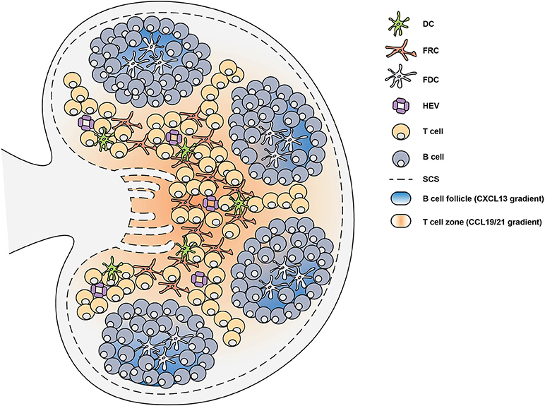
Further, have a look at this cartoon to help understand the locations of the B-cells, T-cells and also dendictric cells in a lymph node 

In your Blood lab please do the following ,  All drawings should be done in a circle and use a petri dish to draw the circle. 

1.  Draw a clear, colour drawing of a human blood smear on high power.  Label the cells that you find, including erythrocytes, and WBCs such as lymphocytes, monocytes, neutrophils, eosinophils. 

2.  Draw a clear, colour drawing of a slide of a lymph node and label whatever structures you can.  Draw this under low and medium power. 

3.  Copy this cartoon of the lymph node to familiarize yourself with where the cells are. 


"Cross-section of a lymph node with sections labelled.1) Capsule; 2) Subcapsular sinus; 3) Germinal center; 4) Lymphoid nodule; 5) Trabeculae" 
copyright Benutzer:Gleiberg